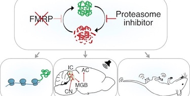
Since its discovery as a strong genetic risk factor for autism, the synaptic scaffolding protein Shank3 has been studied in multiple mouse models. While much of this work has focused on the role of Shank3 in corticostriatal connectivity, our understanding of the impact of Shank3 mutations on large-scale connectivity is far from complete.
Now, SFARI Investigator Alessandro Gozzi and colleagues have shown that homozygous Shank3 mutant mice have robust reductions in long-range and local connectivity in prefrontal cortical areas, which are tightly linked to socio-communicative deficits.
In the new work — partly funded by a SFARI Pilot Award and a previous SFARI Explorer Award — the team used resting state functional magnetic resonance imaging (fMRI) to show long-range desynchronization between prefrontal regions and other long-range cortical targets. They also report desynchronization between the basal ganglia and anterior cingulate cortex and decreased interhemispheric connectivity between striatal regions. These disruptions in connectivity also correlated with observed changes in anogenital sniffing and ultrasonic vocalizations.
All told, the results suggest that Shank3 deficiency leads to selective desynchronization of association and integrative cortical regions, providing another example of an ASD genetic risk factor showing common patterns of prefrontal hypoconnectivity. The data are also broadly consistent with another recent study that implicated the anterior cingulate cortex as a key mediator of the social deficits seen in Shank3 mutant mice (Guo et al., Nat. Neurosci., 2019).
Reference(s)
Deletion of autism risk gene Shank3 disrupts prefrontal connectivity.
Pagani M., Bertero A., Liska A., Galbusera A., Sabbioni M., Barsotti N., Colenbier N., Marinazzo D., Scattoni M.L., Pasqualetti M., Gozzi A.


