Identifying what has gone awry in the brains of individuals with autism is essential for improving diagnosis, and for developing targeted treatments. Converging lines of evidence suggest that the earliest events in brain development play a role in autism. This is supported by observations of autism symptoms in infants.
Several research teams are examining whether and how early development underlies autism. On 12 September, the Simons Foundation Autism Research Initiative brought together a group of scientists to discuss the latest findings on early brain development in autism, based on studies ranging from structural imaging in children to analysis of postmortem brains and mouse models.
In human development, much of the structure of the brain and most of its cells are in place by the beginning of the third trimester of pregnancy.
In the early weeks of the first trimester, brain structure is established and its blueprint, or the patterning of its different regions, begins to form. Brain patterning gives cells important information about their later identity and function. As the brain grows, different cues maintain precisely patterned regions while the cells that make up those regions increase in number.
Cell proliferation extends into the second trimester and allows the cortex of the forebrain to thicken and its surface area to expand. The cortex is the outer layer of the brain and is needed for most higher-order cognitive functions, such as consciousness, memory and attention. Embryonic neural stem cells give rise to neural progenitors, the precursors to neurons. The progenitors then migrate via specific pathways to different regions of the cortex, hippocampus and other brain structures.
The brain then produces glial cells — cells that support neurons — also from specialized progenitor cells. Before birth, these processes are complete. Much of the outgrowth of axons, the long projections of neurons, and dendrites, their signal-receiving branches, depends on the accurate production and placement of their parent cells.
Early structure:
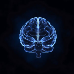
A fundamental neurobiological finding in some people with autism is that their brains are larger than those of controls beginning at an early age. Size differences that are present at birth are likely to be the result of early alterations, which may include changes in the number and organization of cells.
Eric Courchesne, director of the Autism Center of Excellence at the University of California, San Diego, and his collaborators have shown that the front of the cortex is enlarged in postmortem brains from individuals with autism, whereas posterior regions appear to be mostly normal.
This is true for the overall surface area as well as for both gray and white matter. Gray matter consists of the cell bodies of neurons, whereas white matter contains their connecting projections. Changes in brain volume and surface area are associated with language and social skill impairments in children with autism, according to Courchesne.
Flora Vaccarino, professor of neurobiology at Yale University, also highlighted the role of the forebrain and the cortex in the types of brain alterations seen in autism. In mice, dampened signaling through the fibroblast growth factor (FGF) drastically decreases the size of frontal regions of the cortex. Her results show that, in mice, brain size changes are the result of events occurring prior to the outgrowth of axons and dendrites.
Postmortem studies have shown that genes that influence development are expressed differently in the brains of individuals with autism than in controls. The cortex in individuals with autism across a wide range of ages shows disrupted expression of genes that regulate cell number, patterning and differentiation, according to Courchesne. What’s more, children with autism have altered expression of genes important in brain development, such as MAPK, ERK, JAK and STAT. In contrast, older individuals with the disorder show disruptions in repair and signaling pathways.
Daniel Geschwind, director of the Center for Autism Research and Treatment at the University of California, Los Angeles, also identified intriguing links between autism and genes expressed during early development.
His team’s analysis of gene expression in postmortem brains shows that the brains of individuals with autism show less variability across frontal and temporal lobes than control brains do. The researchers also found that autism genes are expressed in neural progenitors derived from stem cells and in fetal human brains, suggesting that they are important for early development.
Geschwind’s team has also found that several genes associated with autism are expressed in specific regions of the developing frontal and temporal cortex. These genes include MET, which codes for an enzyme involved in neural stem cell development and migration, Cadherin 10, an important patterning factor, and CNTNAP2, which regulates the migration of neurons that dampen brain signals.
Patterns and proliferation:
The workshop participants also discussed the genetic, behavioral and structural evidence that implicates two major processes of brain growth in autism: patterning, which determines the types of neurons that form in different brain regions, and neuronal proliferation.
Robert Hevner, professor of neurological surgery at the University of Washington in Seattle, has found that the TBR1 gene, which regulates the expression of other genes, functions as a patterning gene in early development.
Researchers have identified two people with autism who have mutations in TBR1. Embryonic mice lacking TBR1 have lower levels of genes at the front of the brain and higher levels of those at the back of the brain compared with controls.
These findings are consistent with data showing that regions of the front of the brain are dysfunctional in people with autism. Mice lacking TBR1 do not have appropriate distinctions between cell types in the front and the back of the brain, based on gene expression data. They also do not correctly form cortical layers that are key for for sensory and motor functioning. For example, they show a dramatic loss of neurons in the deepest layer, six, and instead are populated with more superficial layer five neurons.
Dennis O’Leary, Vincent J. Coates Chair in Molecular Neurobiology at the Salk Institute in La Jolla, California, described a mechanism that may underlie the patterning deficits seen in autism. Disorders such as autism may result from brain cells developing inappropriate identities that are characteristic of neurons in different regions of the brain. His work has identified genetic factors that operate prenatally to give cells their identities, which develop into areas with distinct functions in the adult cortex.
For example, in embryonic mice lacking EMX2 or COUP-TF1 — regulatory genes highly expressed in the posterior cortex — frontal areas of the cortex expand and posterior areas contract. Also, connections between the cortical areas and the thalamus and other brain regions are altered. These changes result from differences in the expression of these patterning genes and likely contribute to autism, says O’Leary.
Anjen Chenn, associate professor of pathology at the University of Illinois in Chicago, reported that beta-catenin — a molecule that relays signals through the Wnt pathway — regulates the proliferation of neural progenitors in the cortex.
A large sequencing study published in April found that spontaneous, or de novo, mutations in beta-catenin are more common in individuals with autism than in controls. What’s more, nearly 40 percent of the de novo mutations identified in people with autism are also involved in the Wnt pathway.
In mice engineered to express elevated levels of beta-catenin, neural progenitors are in an actively dividing state for longer than they are in controls. This results in a larger cortex with a more convoluted surface area than is seen in controls. The results for both beta-catenin and FGF suggest that core developmental pathways that influence progenitor cells in the cortex may underlie abnormalities in autism.
The researchers also showed links between autism mouse models and genes involved in early development. For example, genes encompassed by the 22q11 and 16p11.2 copy number variants, both of which involve developmental disabilities, regulate signaling through the ERK pathway.
Mouse models of 22q11 deletion syndrome make fewer precursors of excitatory neurons than controls do, says Anthony-Samuel LaMantia, director of George Washington University’s Institute for Neuroscience in Washington, D.C. Neurons that signal through gamma-aminobutyric acid, which inhibits brain signals, are seen at different sites in the cortex than they are in controls. These deficits are most pronounced in the frontal cortex.
The mice also have problems with working memory, which is regulated by the frontal cortex. Deletion of the 22q11 chromosomal region results in diminished expression of the CXCR4 gene (which is located outside the deleted region). CXCR4 is involved in the development of inhibitory neurons in the brain.
Mice lacking the ERK2 gene and those with deletions within 16p11.2 show disruptions in the proliferation of neural progenitor cells early in embryogenesis. These mice are also more anxious and have more memory problems than controls do, says Gary Landreth, director of the Alzheimer Research Laboratory at Case Western Reserve University in Cleveland, Ohio. His work links embryonic production of neurons to autism symptoms in the mice.
The multiple approaches presented at the workshop all implicate early developmental processes in autism. However, the investigators emphasized that mouse models can only suggest the mechanisms at play in people. For example, mice lacking the FGF receptor have a drastically smaller cortex than controls do, whereas people with autism show only small alterations in cortical volume.
The participants also discussed why some individuals with the disorder have severe behavioral problems but subtle neurobiological changes. They acknowledged that although many people with autism are severely impaired, they have good basic brain function needed for motor control, eating and sleeping and sometimes for complex skills such as music, reading and learning. This is consistent with the mostly intact brain structures found in people with autism.
To explore these findings further, researchers will need sophisticated genetic techniques. Because mouse models have inherent limitations, the investigators discussed other methods, particularly using human cells. For example, induced pluripotent stem cells made from people with autism, along with fine-grained analyses across developmental stages of the human embryonic brain, may identify the disorder’s origins in neural patterning or proliferation. They may also be able to suggest how to put development back on track.


