Loss of the autism risk gene PTCHD1 disrupts thalamic reticular function and results in attention deficits in mice
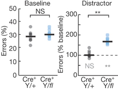
Attention deficits: Mice lacking PTCHD1 in the TRN (Cre+ Y/fl) display similar baseline performance in a visual detection task compared with control mice (Cre+ Y/+) but have decreased accuracy in the presence of distractors. Dashed line, baseline performance (without distractors). Image from Wells M.F. et al. (2016).Mutations in the patched domain containing 1 (PTCHD1) gene have been identified in about 1 percent of individuals with autism spectrum disorder (ASD) and intellectual disability. These individuals may display a range of symptoms, including attention deficits, hyperactivity, sleep abnormalities and learning disabilities. SFARI Investigators Guoping Feng and Michael Halassa have now developed genetic mouse models that help to explain how loss of PTCHD1 within specific brain circuitry may cause particular ASD phenotypes. The researchers have found that PTCHD1 is selectively expressed in the thalamic reticular nucleus (TRN) of mice, a brain region that regulates sensorimotor processing, attention and the generation of sleep rhythms. By creating two strains of mice with either a global deletion of PTCHD1 or specific deletion in the TRN, Feng and Halassa’s teams demonstrate that select behavioral phenotypes — namely, attention deficits and hyperactivity — are specifically caused by TRN dysfunction. Moreover, these defects can be rescued by pharmacological augmentation of small conductance calcium-dependent potassium (SK) channel activity. Such findings not only enhance our understanding of specific neural circuits that are responsible for clinically relevant behaviors, but also identify a potential pharmacological target for treating ASD symptoms.
Reference(s)
Thalamic reticular impairment underlies attention deficit in PTCHD1Y/- mice.