A role for the maternal interleukin-17A pathway in autism
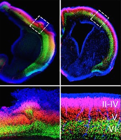
Cortical development gone awry: Patches of disorganized cortex were observed in fetal brains derived from MIA pregnant mice (left panels). Pretreatment with antibodies that block IL-17A activity resulted in normal cortical development (right panels). Image from Choi G.B. et al. (2016).Previous studies have found an association between maternal infection and autism spectrum disorder (ASD) in the child. SFARI Investigator Dan Littman and his collaborators (including Simons Center for the Social Brain Investigators Gloria Choi and Yeong Yim) used a mouse maternal immune activation (MIA) model to further dissect which immune cells and pathways are involved in this association. They found that maternal interleukin-17A, a cytokine mediator of T helper 17 (TH17) cells, induces abnormal cortical development — a finding that has previously been observed in genetic mouse models of ASD and postmortem tissue from people with ASD — and ASD-like behavioral phenotypes in offspring of MIA mice. Importantly, treatment of pregnant mice with antibodies that block IL-17A activity partially protected against the development of MIA-induced behavioral abnormalities. These findings suggest that modulating the TH17/interleukin-17A pathway may represent a novel therapeutic approach for reducing prenatal infection-induced inflammation that can contribute to the development of ASD.
Reference(s)
The maternal interleukin-17a pathway in mice promotes autism-like phenotypes in offspring.