The brain is the main organ affected in autism spectrum disorder (ASD), yet little is known about the neuropathological changes linked to the condition. To facilitate stereological analyses of autism postmortem brains, Autism BrainNet is now supporting an effort to digitize the Autism Celloidin Library.
Started in the early 2000s under the auspices of Autism Speaks as part of the Autism Tissue Program (now managed by Autism BrainNet), the Autism Celloidin Library comprises 28 postmortem brains from 14 donors with ASD and 14 neurotypical individuals, ranging from 4 to 50 years old. The brains in the collection have been embedded in 8 percent celloidin, with one hemisphere being cut into serial 200-μm-thick sections. All sections are mounted on histological slides, which have been stained with cresyl violet. This resource, which is now part of the Autism BrainNet brain collection, is housed at the Icahn School of Medicine at Mount Sinai. Autism BrainNet collaborator Patrick Hof is leading the digitalization process one slide at a time.
“The Autism Celloidin Library is a unique resource for exploratory studies as well as large-scale histopathological investigations. These studies are instrumental to understanding cell population-specific changes in the brains of individuals with ASD,” says Hof.
By creating a digital repository of the collection, Autism BrainNet aims to make this resource widely accessible to interested researchers and to create a permanent digital tissue record. This will ensure a broad use of the collection for research studies without risking compromising the quality and integrity of the specimens, which are mounted on large glass slides and are, therefore, extremely fragile.
To maximize the value of the collection for the scientific community, Hof also plans to anatomically annotate the digitized slides. This will provide a cytoarchitectonic guide to researchers and help them navigate the cellular organization of the tissue with sufficient detail and resolution for conducting high-quality quantitative analyses.
To date, over 20 published studies have used the Autism Celloidin Library. This research has provided insights into neuropathological differences in neuron number, density and area volume across several cortical and subcortical regions in the brain of people with ASD (e.g., 1-9).

“We are excited to digitize this collection,” says Marta Benedetti, SFARI senior scientist and Autism BrainNet managing director. “Our goal is to provide an easy-to-access resource to study the cellular neuropathology of ASD, while ensuring the conservation of these unique materials for the years to come.”
Images from the collection will be made available for research purposes in Spring 2022.
Interested researchers can contact [email protected] for further information.
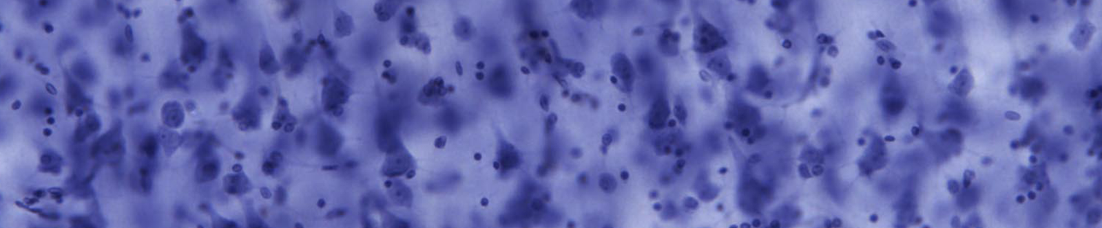
Amygdala view. Close-up images of the amygdala and its cellular structure (40x magnification) from Nissl-stained slides of the Autism Celloidin Library. Images obtained from Patrick Hof/Icahn School of Medicine at Mount Sinai
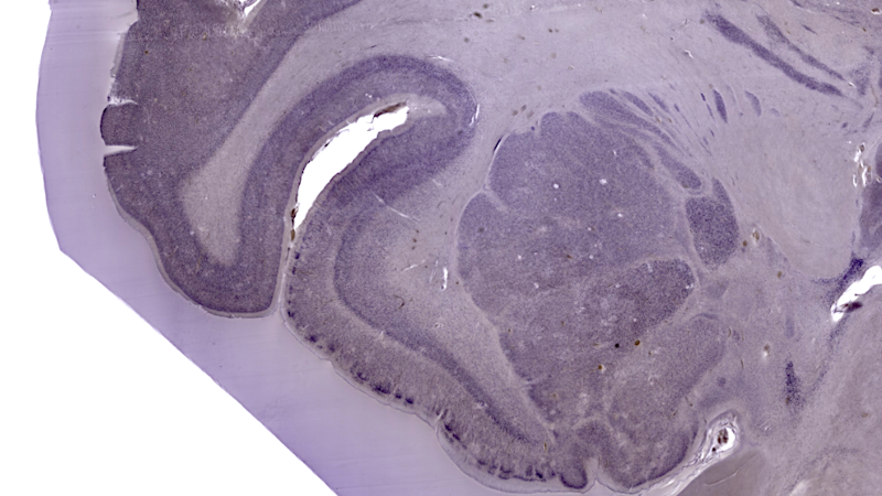
Amygdala view. Close-up images of the amygdala and its cellular structure (40x magnification) from Nissl-stained slides of the Autism Celloidin Library. Images obtained from Patrick Hof/Icahn School of Medicine at Mount Sinai
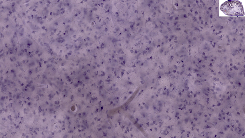
Amygdala view. Close-up images of the amygdala and its cellular structure (40x magnification) from Nissl-stained slides of the Autism Celloidin Library. Images obtained from Patrick Hof/Icahn School of Medicine at Mount Sinai
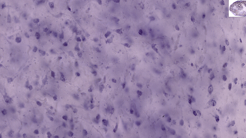
Amygdala view. Close-up images of the amygdala and its cellular structure (40x magnification) from Nissl-stained slides of the Autism Celloidin Library. Images obtained from Patrick Hof/Icahn School of Medicine at Mount Sinai
References
- Van Kooten I. A. J. et al. Brain 131, 987-999 (2008) PubMed
- Santos M. et al. Brain Res. 1360, 206-217 (2011) PubMed
- Jacot-Descombes S. et al. Acta Neuropathol. 124, 67-79 (2012) PubMed
- Uppal N. et al. J. Neuropathol. Exp. Neurol. 73, 891-902 (2014) PubMed
- Wegiel J. et al. Acta Neuropathol. Commun. 2, 28 (2014) PubMed
- Uppal N. et al. Mol. Autism 5, 17 (2014) PubMed
- Wegiel J. et al. Acta Neuropathol. Commun. 3, 2 (2015) PubMed
- Wegiel J. et al. Acta Neuropathol. Commun. 2, 141 (2014) PubMed
- Avino T.A. et al. Proc. Natl. Acad. Sci. USA 115, 3710-3715 (2018) PubMed


